Seeing The Brain In 3D
- Oct 1, 2025
- 3 min read
Updated: Jan 23
A GPS for the Brain: How Mixed Reality Is Changing Neurosurgery

How Surgeons Navigate the Brain
When we think of surgery, we often picture precision scalpels and steady hands. But in modern neurosurgery, one of the most important “instruments” isn’t in the surgeon’s hand, it’s in front of their eyes. Navigation systems have become the brain’s GPS: they fuse MRI and CT images into 3D maps that guide the surgeon through the most complex and delicate terrain in the human body.
These systems translate medical imaging into a real-time map of the brain, allowing doctors to locate a tumor, avoid critical blood vessels or motor areas, and minimize healthy tissue damage. The result? Shorter operations, smaller incisions, and greater safety.
Yet traditional optical navigation equipment is bulky, expensive, and confined to large hospitals—limiting access to cutting-edge precision.
The Rise of Mixed Reality in the Operating Room
To overcome these limits, researchers are now experimenting with mixed reality (MR), a technology that blends virtual and physical space through head-mounted displays such as Microsoft’s HoloLens 2. Imagine wearing a visor that lets you see both the patient and a holographic model of their brain, aligned perfectly in 3D.
The surgeon no longer needs to glance between a monitor and the patient’s head; the virtual brain is literally projected onto reality.
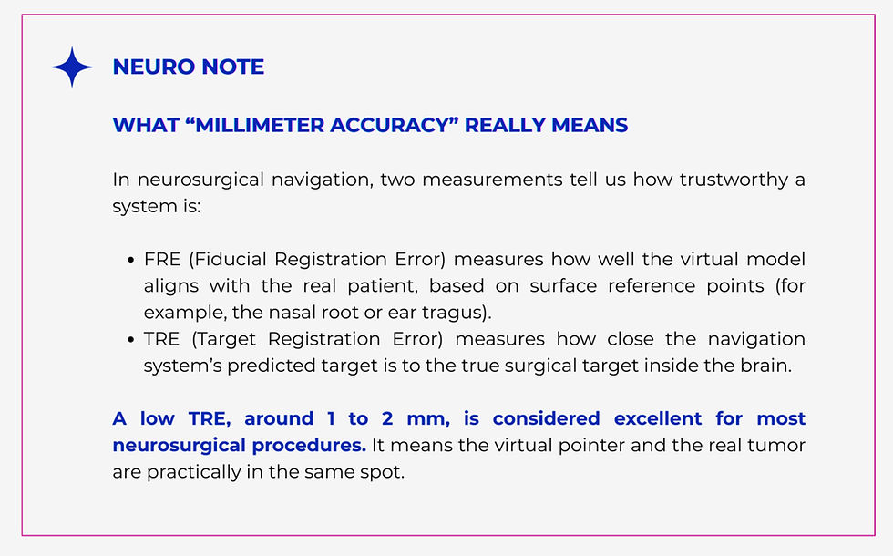
A new study introduced a Portable Mixed Reality Navigation (PMRN) system designed to make this technology accessible, affordable, and accurate. The system connects a headset and a laptop via a simple local router. Using infrared tracking, it localizes a surgical probe in real time and overlays the patient’s anatomy with millimeter precision.
Researchers tested PMRN first in simulation labs, then during surgeries on 42 patients with intracranial lesions.
The results were striking: the average localization error was about 1.7 mm, with 95% surgeon satisfaction. Setup time dropped as doctors practiced, stabilizing at around 4 minutes per case.
For many operations, that level of precision is more than enough.

How It Works: Seeing Through the Skull
The PMRN system builds a 3D model from the patient’s MRI or CT scans. Once the surgeon wears the headset, the holographic brain is projected onto the actual head, locked in space using spatial tracking algorithms (called SLAM).
To reduce errors in depth perception, a common problem when looking at 3D objects through a single pair of lenses, the researchers added auxiliary panels that show synchronized 2D views of the surgical probe from different angles. The surgeon can also use voice commands to interact with the system, keeping their hands sterile and focused.
The experience feels like operating “through” the skull, with both real and virtual anatomy visible at once. Surgeons described the alignment between hologram and patient as “natural” and “intuitive,” noting that they quickly adapted to the new visual field.
Precision, but with Limits
The PMRN system proved accurate enough for most routine brain surgeries, such as hematoma evacuation, ventricular drainage, or tumor resections (like meningiomas).
But procedures requiring sub-millimeter precision, such as deep brain stimulation or needle biopsies in deep structures, still need ultra-refined systems.
Another challenge is the “brain shift”: after the skull is opened, cerebrospinal fluid release causes the brain to subtly move. Since PMRN relies on preoperative imaging, any small anatomical shift can slightly distort accuracy. Researchers suggest combining MR with intraoperative ultrasound, which updates the anatomy in real time.
Why This Matters for the Next Story
This technology captures the essence of what associations like ASINO Foundation for Oncological Neurosurgery support: making advanced neurosurgical tools and training accessible. Behind every technical improvement lies the goal of saving neurons, protecting functions, and giving patients safer outcomes.
Conclusion
Mixed Reality is redefining how surgeons see and navigate the brain, turning complex anatomy into an intuitive, shared visual space.
By making precision more accessible, this technology doesn’t just improve surgery,
it brings us closer to a future
where innovation truly serves patient safety and care.
Source:
Zhou, Y. et al. (2023). A portable mixed reality navigation system for neurosurgery.
Frontiers in Neuroscience. Microsoft HoloLens 2 – Medical Mixed Reality Applications, clinical and technical documentation. Cutting, C. et al. (2021).
Augmented and mixed reality in neurosurgery: current applications and future perspectives. Neurosurgical Review.


Comments